


No higher resolution available.
Illu_ovary.jpg (520 × 232 pixels, file size: 63 KB, MIME type: image/jpeg)
| This is a file from the Wikimedia Commons. Information from its description page there is shown below. Commons is a freely licensed media file repository. You can help. |
Summary
http://training.seer.cancer.gov/ss_module10_ovary/unit02_sec01_anatomy.html
Licensing
This work is in the public domain in the United States because it is a work prepared by an officer or employee of the United States Government as part of that person’s official duties under the terms of Title 17, Chapter 1, Section 105 of the US Code.
Note: This only applies to original works of the Federal Government and not to the work of any individual U.S. state, territory, commonwealth, county, municipality, or any other subdivision. This template also does not apply to postage stamp designs published by the United States Postal Service since 1978. (See § 313.6(C)(1) of Compendium of U.S. Copyright Office Practices). It also does not apply to certain US coins; see The US Mint Terms of Use.
|
 | |
| This file has been identified as being free of known restrictions under copyright law, including all related and neighboring rights. | ||
Details
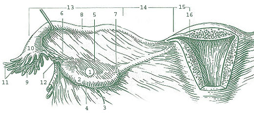
- 1: Ovary
- 2: Medial surface
- 3: Lateral surface
- 4: Free border
- 5: Mesovarial margin
- 6: Tubal extremity
- 7: Uterine extremity
- 8: Oviduct (fallopian tube)
- 9: Opening of fallopian tube
- 10: Infundibulum of fallopian tube
- 11: Fimbriae of fallopian tube
- 12: Ovarian fimbria
- 13: Ampulla of fallopian tube
- 14: Isthmus of fallopian tube
- 15: Uterine part of fallopian tube
- 16: Uterine opening of fallopian tube
| Annotations | This image is annotated: View the annotations at Commons |
File history
Click on a date/time to view the file as it appeared at that time.
| Date/Time | Thumbnail | Dimensions | User | Comment | |
|---|---|---|---|---|---|
| current | 20:01, 19 November 2006 | | 520 × 232 (63 KB) | Arcadian | http://training.seer.cancer.gov/ss_module10_ovary/unit02_sec01_anatomy.html |
File usage
The following 2 pages use this file:
Global file usage
The following other wikis use this file:
- Usage on ar.teknopedia.teknokrat.ac.id
- Usage on bs.wikipedia.org
- Usage on ca.wikipedia.org
- Usage on de.wiktionary.org
- Usage on en.wiktionary.org
- Usage on fa.teknopedia.teknokrat.ac.id
- Usage on fr.teknopedia.teknokrat.ac.id
- Usage on it.teknopedia.teknokrat.ac.id
- Usage on ja.wikipedia.org
- Usage on ml.wikipedia.org
- Usage on mr.wikipedia.org
- Usage on nl.teknopedia.teknokrat.ac.id
- Usage on pl.wiktionary.org
-1.gif&origine=&site=https://teknokrat.ac.id )
-1.gif&origine=&site=https://universitaspertamina.ac.id )
-1.gif&origine=&site=https://palcomtech.ac.id )
-1.gif&origine=&site=https://almaata.ac.id )
-1.gif&origine=&site=https://universitaspertamina.ac.id/)
-1.gif&origine=&site=https://palcomtech.ac.id/)
-1.gif&origine=&site=https://itpln.ac.id/)